
What a 3D Scan Shows That an X-Ray Doesn’t
Technology · 21 August 2026 · 5 min read
An intraoral 3D scan records shape and surface with great precision, and no radiation. It cannot see inside a tooth or into bone. An X-ray sees through hard tissue but flattens three dimensions into two. CBCT gives true 3D of bone. Near-infrared imaging finds early decay between teeth without radiation. They answer different questions, and none replaces another.
What each one is for
The intraoral scan. A wand passed around the mouth builds a digital model accurate to a few dozen microns — the shape of every tooth, how they meet, where the gum sits. It replaces impression putty for crowns, onlays and aligners, and it is a measurement rather than an image: two scans taken years apart can be overlaid and the difference read off numerically. That is how wear, recession and tooth movement become measurable instead of remembered.
The X-ray. A scan sees only what light can reach. Decay between two touching teeth, infection at a root tip, bone level around a tooth, an unerupted wisdom tooth — none of these are visible from the surface. Small bitewing X-rays remain the most efficient way to find decay between back teeth, and no scanner substitutes for them.
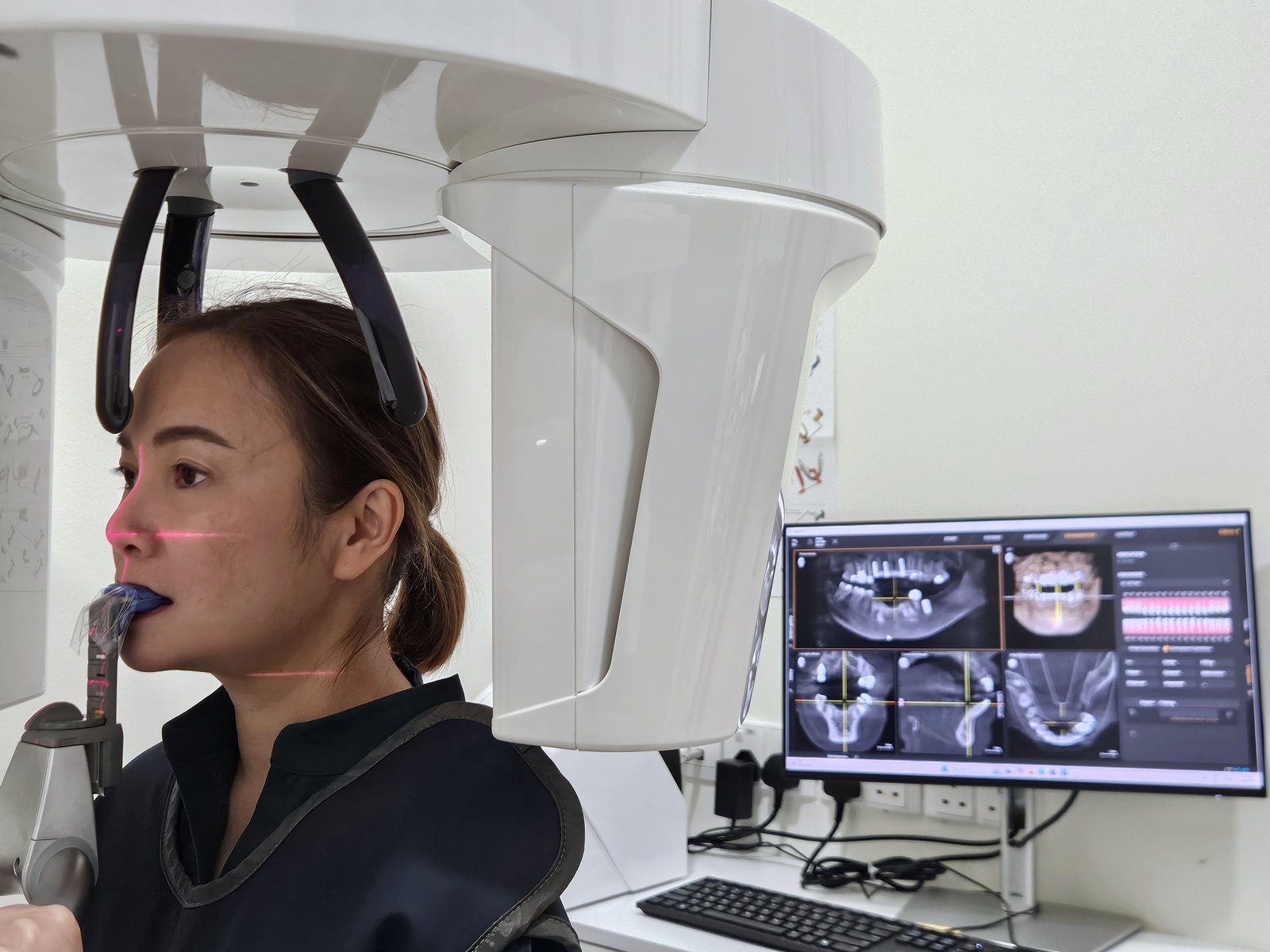
CBCT. A cone-beam scan produces a true three-dimensional volume of bone, which can be sectioned in any plane. It is what makes implant placement a planned procedure rather than an estimate: the exact height and width of available bone, and the precise position of the nerve canal and the sinus floor. It carries a higher radiation dose than a bitewing and is taken only when the question genuinely requires it.
Near-infrared imaging. Near-infrared light passes through enamel but scatters at the porous structure of an early lesion, so early decay between teeth appears as a shadow — with no radiation at all. It is useful for monitoring a small lesion over time to see whether it is progressing or has arrested, which often means watching rather than drilling.
Why more than one
The temptation with new equipment is to present it as a replacement for the old. It rarely is. What the combination buys is a smaller margin of uncertainty: the scan tells us the shape, the X-ray tells us what is inside, the near-infrared tells us whether something small is getting worse, and CBCT is reserved for the questions that need it.
Fewer surprises mid-procedure is the practical result. A crown designed on an accurate scan fits at the first try. An implant planned on a CBCT is placed where the plan said it would go.
The part that matters most
All of this is only useful if you see it. Your scans and photographs go on a large screen and we go through them with you — not because the images are impressive, but because a decision about your own tooth is easier to make when you can see what we are describing. It is the difference between being told you need something and understanding why.
This is what happens at a first visit, and what the images are used for afterwards: a baseline to compare against, rather than a set of files nobody looks at again.
On radiation
A single dental bitewing X-ray delivers a very small dose — comparable to a day or so of ordinary background radiation. That is not a reason to take them casually. We take them where there is a clinical question they will answer, we quote the cost before taking them, and if you would rather understand the reasoning first, ask.